Attic Cholesteatoma Radiopaedia

Cholesteatoma Radiology Reference Article Radiopaedia Org

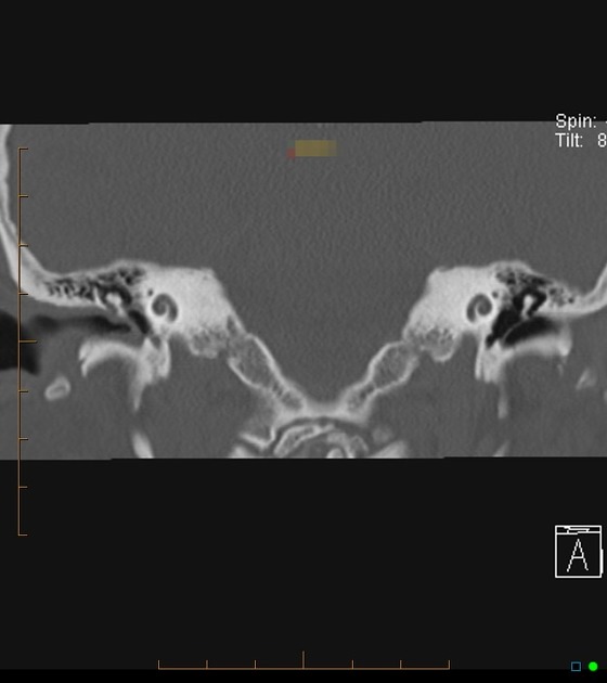
Fd Acquired Pars Flaccida Cholesteatoma Left Coronal T Bone Ct Image Shows An Atticoantral Nondependent Homogeneous Soft Radiology Image Shows Head And Neck

Mastoditis Middle Ear Head And Neck Sinusitis

Cholesteatoma Radiology Case Radiopaedia Org
Acquired Cholesteatoma Radiology Reference Article Radiopaedia Org

Cholesteatoma Radiology Case Radiopaedia Org
The overall incidence rate in one large study was 0 30 per year per 100 000 inhabitants 1.
Attic cholesteatoma radiopaedia.

Image Result For Scutum Erosion Facial Nerve Eustachian Tube Dysfunction Middle Ear

Mastoditis Middle Ear Head And Neck Sinusitis
Pars Tensa Cholesteatoma Radiology Case Radiopaedia Org

Ct Through The Orbits Obtained Initially Without Contrast And Then With Contrast While The Patient Performed A Valsalva Manoeuvre In The Kt Ppn

Huge Cholesteatoma Radiology Mural Ear

Cholesteatoma Acquired Radiology Case Radiopaedia Org Radiology Sonography Case

The Radiology Assistant Pathology

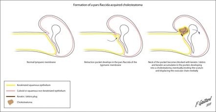
Formation Of A Cholesteatoma Radiology Case Radiopaedia Org Medical Illustration Radiology Middle Ear

The Radiology Assistant Temporal Bone Anatomy 2 0
Tympanic Membrane Retraction Radiology Reference Article Radiopaedia Org

Paediatric Cholesteatoma Eurorad

Middle Ear And Mastoid Cholesteatoma Postoperative View The Primary Goal Of Surgery For Chronic Otitis Media And Cholesteatoma Otitis Media Otitis Middle Ear

Female Reproductive System Radiology Key Radiology Female Reproductive System Reproductive System Female

Mondini Malformation Radiology Reference Article Radiopaedia Org Radiology Pet Ct Head And Neck
Source : pinterest.com
